From the scanner
One phantom, six contrasts
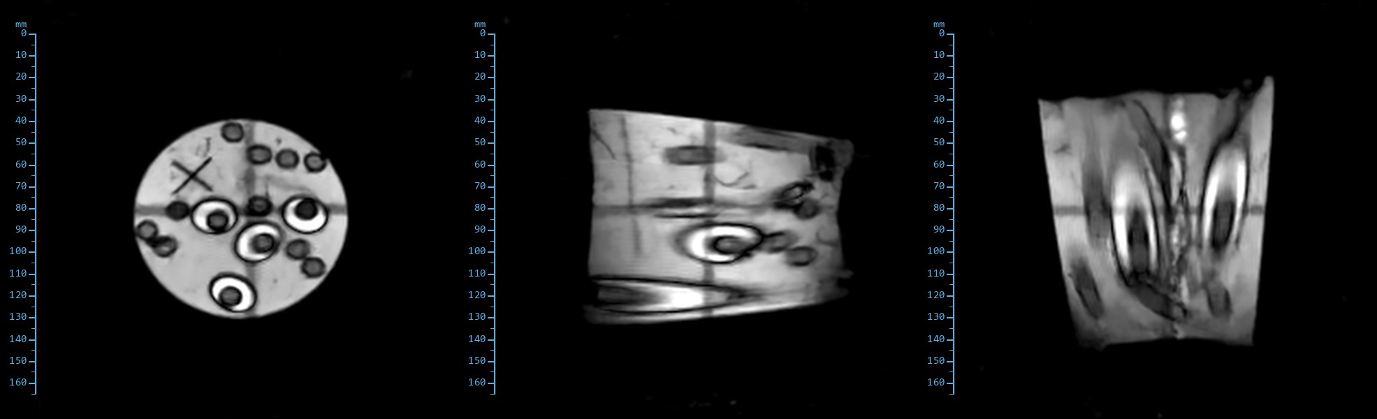
Acquired on the AltraMRI 0.4T system on 9 November 2026. A three-media phantom run through the standard fore-foot protocol: pilot, proton density, T1, T2, T2* and STIR, twelve 5 mm slices each, 170 mm field of view, 1 mm in-plane voxels. Rendered from the console's reconstructions, windowed per image, with a millimetre scale on each.
The phantom
Three media, three tissues
A disc of doped agar carrying tubes of pure water and MCT oil. Each medium stands in for a tissue the fore-foot protocol has to separate.
Pure water
The small round tubes
Free fluid: joint effusion, a cyst, oedema
Very long T1 (seconds) and long T2. Saturated and dark whenever TR is short; the brightest thing in the phantom on T2 and STIR.
MCT oil
The larger tubes around some of the water tubes
Fat: the digital cushion, marrow
Short T1, so bright on every T1-weighted image. Its inversion time of roughly 90 ms matches fat in the equine limb at 0.4T, so the STIR nulls it.
Agar, copper sulphate, salt
The bulk of the disc
Muscle and soft tissue
Doped to the T1 and T2 of muscle. Recovers between repetitions, so intermediate grey on T1 and proton density, and darker than fluid on T2.
Same slice, six sequences
How the three media rank
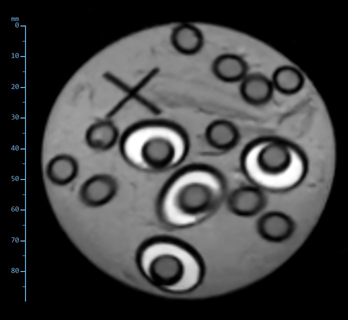
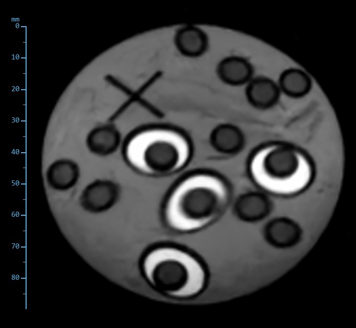
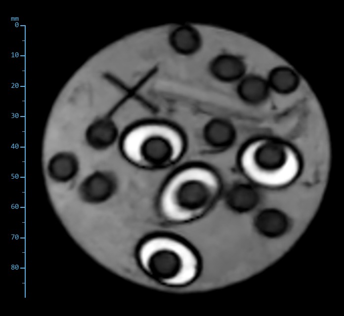
Slice 5 of 12 from each scan, cropped to the disc. Read the water tubes first, then the oil rings, then the agar between them.
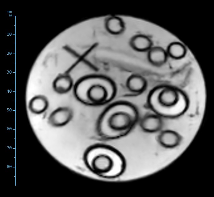
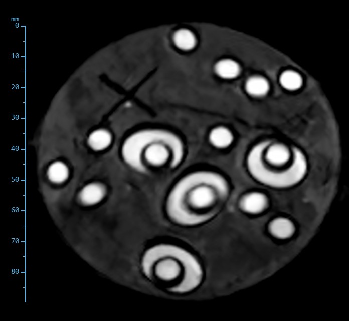
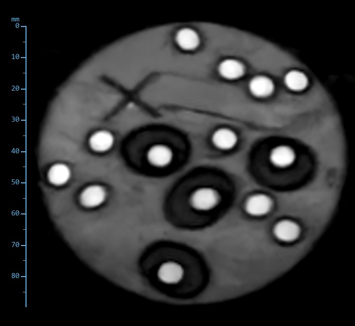
| Sequence | TR / TE | Acquired matrix, voxel | Water (fluid) | MCT oil (fat) | Doped agar (muscle) |
|---|---|---|---|---|---|
| Proton density Spin echo | 1000 / 24 ms | 170 × 170, 1.0 × 1.0 × 5 mm | dark | bright | intermediate |
| T1-weighted Spin echo | 480 / 20 ms | 170 × 170, 1.0 × 1.0 × 5 mm | dark | bright | intermediate |
| T1-weighted Gradient echo | 116 / 9 ms | 170 × 170, 1.0 × 1.0 × 5 mm | dark | bright | intermediate |
| T2*-weighted Gradient echo | 136 / 9 ms | 170 × 170, 1.0 × 1.0 × 5 mm | intermediate | intermediate | bright |
| T2-weighted Fast spin echo | 2000 / 84 ms | 168 × 180, 1.0 × 0.9 × 5 mm | bright | bright | dark |
| Fluid-sensitive, fat-suppressed STIR fast spin echo | 3160 / 28 ms | 168 × 180, 1.0 × 0.9 × 5 mm | bright | nulled | intermediate |
What it shows
Three things to take from the phantom
The ordering is right
Across six sequences the three media rank exactly as fluid, fat and muscle rank in the limb. That is the test a contrast phantom is built for.
STIR separates fluid from fat
With oil at the inversion null and water at its brightest, the fat-suppressed image is the one that will show bone oedema against marrow fat.
Spin echo and gradient echo behave as they should
Spin echo refocuses the susceptibility at every tube wall; gradient echo does not and draws the rings. Both families are available in every plane.
Through the stack
Twelve slices, two of the sequences
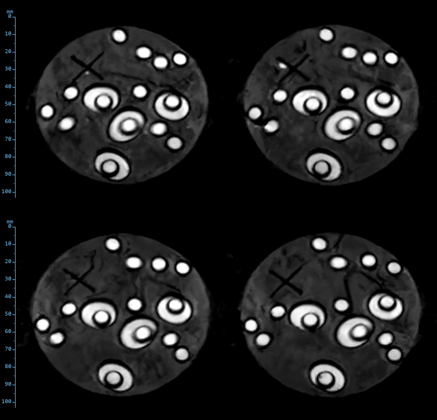
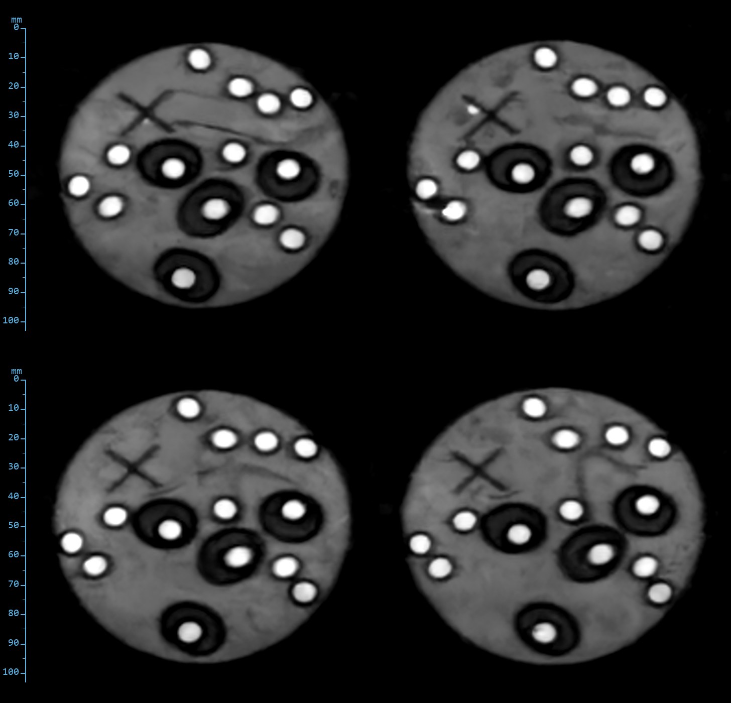
Reconstructions from the AltraMRI console, 9 November 2026, windowed per image; standard reconstruction, no further processing. Scales in millimetres from the acquisition field of view. Matrices are as acquired: readout oversampling and navigator echoes are not counted. Phantom images; no horse has been imaged on the 0.4T system yet. The phantom was first imaged through the full protocol on 8 November 2026 and re-acquired on 9 November with the standard fast spin echo readout.