From the grocery store

Seven things from the shop, six contrasts

Acquired on the AltraMRI 0.4T system on 8 and 9 November 2026. A pork hock, a grapefruit, a pomegranate, three duck eggs, three kiwis, an avocado and a bell pepper, each run through the same fore-foot protocol as the phantom: pilot, proton density, T1, T2, T2* and STIR, twelve 5 mm slices, 170 mm field of view, 1 mm in-plane voxels. Nothing was changed between objects except the object.
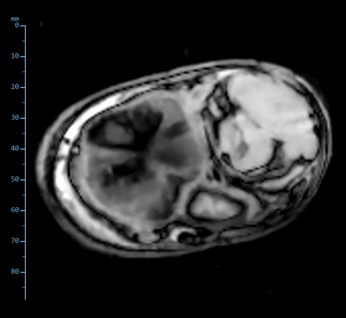
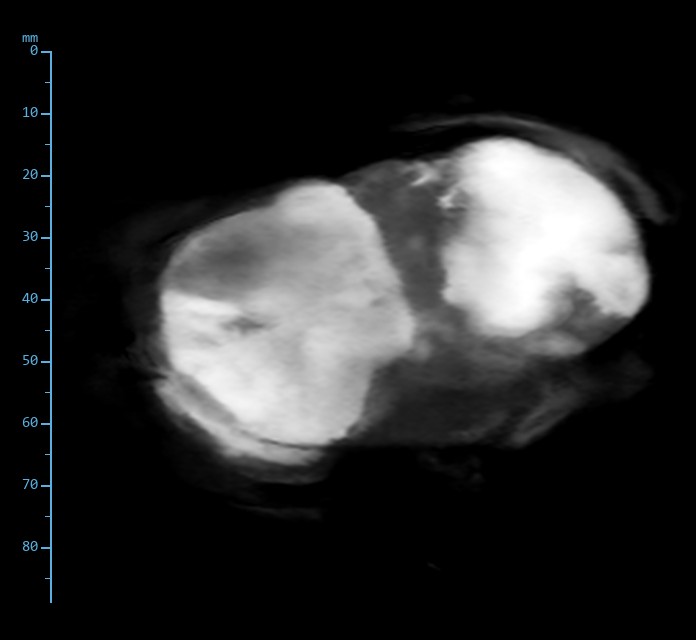
Pork hock, T2 FSE
Pork hock, T2 FSE
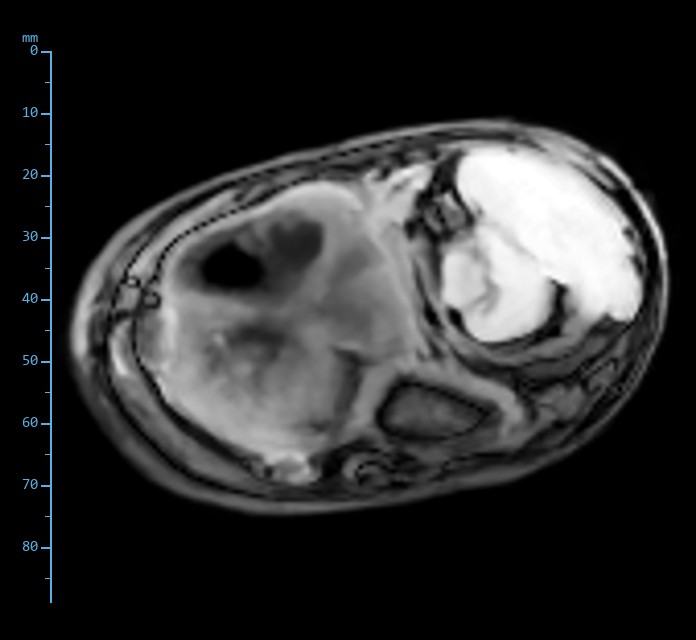
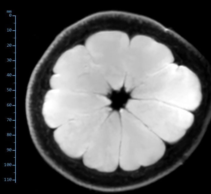
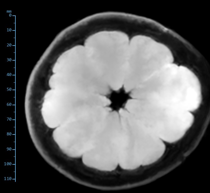
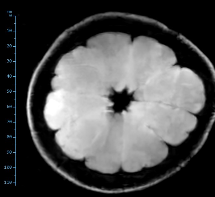
Grapefruit, T2 FSE
Grapefruit, T2 FSE
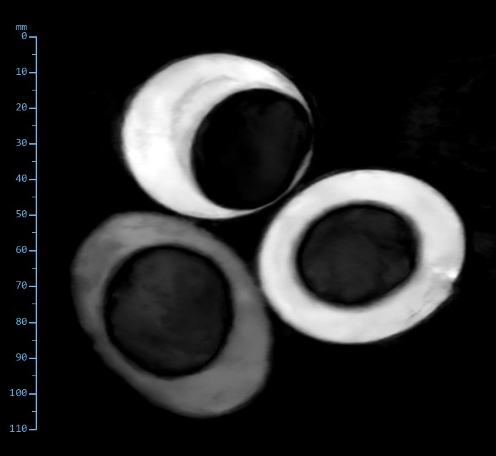
Duck eggs, STIR
Duck eggs, STIR
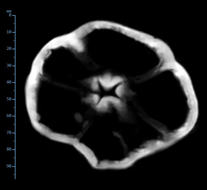
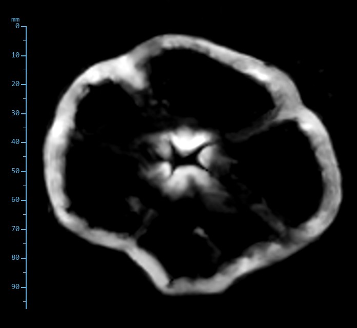
Bell pepper, T2* GRE

Meat counter

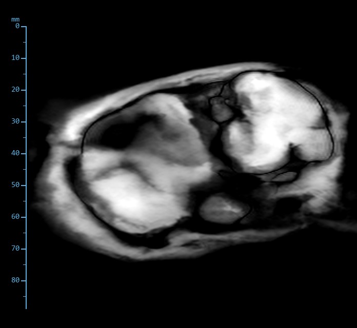
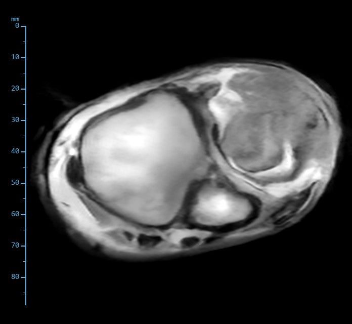
Pork hock: Skin, fat, muscle, tendon, bone

A transverse slice through a pork hock: the closest thing to a limb that a supermarket sells. Read it as you would a horse: skin and subcutaneous fat at the rim, muscle bellies and tendons around two joint ends, cortical bone black on everything.

Slice 11 of 12. Same slice, six sequences.
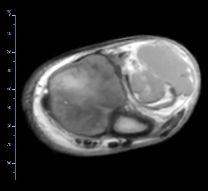
Proton density spin echo image of the pork hock
Proton density Spin echoTR 1000 ms · TE 24 ms · 170 × 170, 1.0 × 1.0 × 5 mmSubcutaneous fat bright, muscle intermediate, tendons and cortical bone black. The large joint end is intermediate: dark on T1 and bright on T2, it reads as cartilage rather than fatty marrow, as expected in a market-age pig.
T1-weighted spin echo image of the pork hock
T1-weighted Spin echoTR 480 ms · TE 20 ms · 170 × 170, 1.0 × 1.0 × 5 mmFat brightest, muscle grey, the cartilaginous joint end dark. The classic T1 anatomy picture.
T1-weighted gradient echo image of the pork hock
T1-weighted Gradient echoTR 116 ms · TE 9 ms · 170 × 170, 1.0 × 1.0 × 5 mmSame ordering as the spin echo in a quarter of the time. Fine dark lines mark the fascial planes.
T2*-weighted gradient echo image of the pork hock
T2*-weighted Gradient echoTR 136 ms · TE 9 ms · 170 × 170, 1.0 × 1.0 × 5 mmSusceptibility draws a sharp black line at every cortical bone surface and tendon edge.
T2-weighted fast spin echo image of the pork hock
T2-weighted Fast spin echoTR 2000 ms · TE 84 ms · 168 × 180, 1.0 × 0.9 × 5 mmThe cartilaginous joint end lights up, as a joint effusion would. Fat stays bright on fast spin echo, muscle falls away.
STIR fast spin echo, ti 95 ms image of the pork hock
STIR Fast spin echo, TI 95 msTR 3160 ms · TE 28 ms · 168 × 180, 1.0 × 0.9 × 5 mmFat suppressed, the water-rich joint end brightest. This is the contrast that finds fluid in and around bone.
T2-weighted fast spin echo: four slices through the pork hock
T2-weighted fast spin echo. Slices 7, 9, 10 and 11 of 12. TR 2000 ms, effective TE 84 ms, 168 × 180 acquired, 1.0 × 0.9 × 5 mm.

Produce

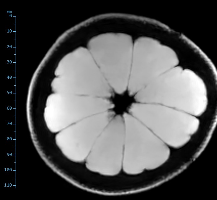
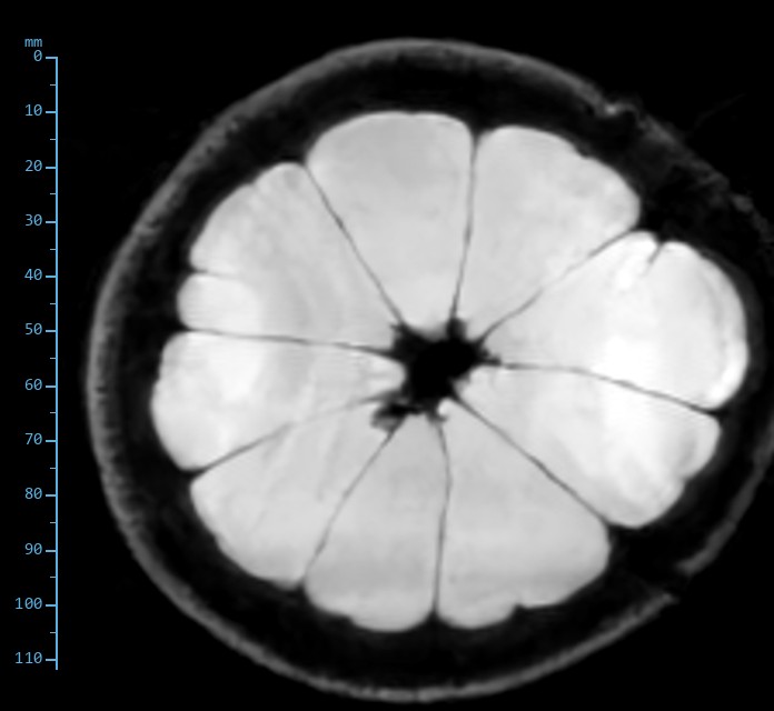
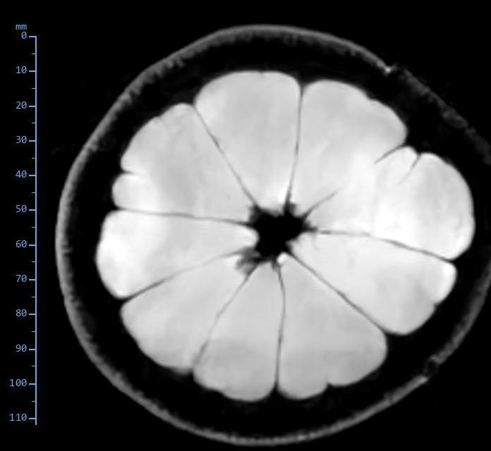
Grapefruit: Membranes one millimetre thick

The MRI textbook fruit. Juice vesicles are almost pure water, the segment membranes and pith are nearly dry, and the peel carries oil glands. The question it answers is resolution: can the septa be followed from core to rind?

Slice 3 of 12. Same slice, six sequences.

Proton density spin echo image of the grapefruit
Proton density Spin echoTR 1000 ms · TE 24 ms · 170 × 170, 1.0 × 1.0 × 5 mmSegments bright, septa dark, pith intermediate, peel a thin bright rim.
T1-weighted spin echo image of the grapefruit
T1-weighted Spin echoTR 480 ms · TE 20 ms · 170 × 170, 1.0 × 1.0 × 5 mmPith brightens relative to the segments; the peel is the brightest ring.
T1-weighted gradient echo image of the grapefruit
T1-weighted Gradient echoTR 116 ms · TE 9 ms · 170 × 170, 1.0 × 1.0 × 5 mmLower matrix, the same picture.
T2*-weighted gradient echo image of the grapefruit
T2*-weighted Gradient echoTR 136 ms · TE 9 ms · 170 × 170, 1.0 × 1.0 × 5 mmHighest contrast of the set: septa and the air-filled core black against bright segments.
T2-weighted fast spin echo image of the grapefruit
T2-weighted Fast spin echoTR 2000 ms · TE 84 ms · 168 × 180, 1.0 × 0.9 × 5 mmLong T2 everywhere water is; the dry pith drops out and the septa stay crisp.
STIR fast spin echo, ti 95 ms image of the grapefruit
STIR Fast spin echo, TI 95 msTR 3160 ms · TE 28 ms · 168 × 180, 1.0 × 0.9 × 5 mmThe peel oil nulls. Everything else behaves as it does on T2.
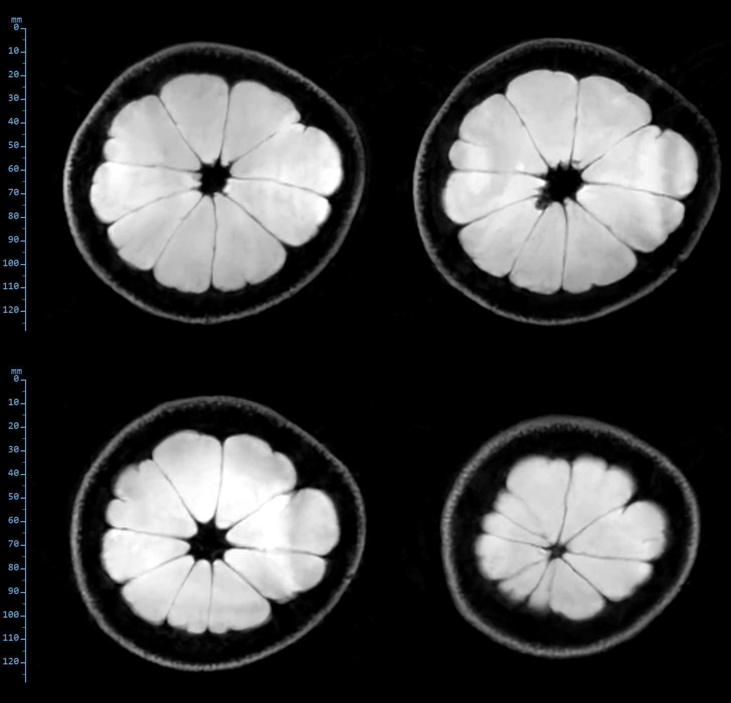
T2-weighted fast spin echo: four slices through the grapefruit
T2-weighted fast spin echo. Slices 2, 4, 7 and 10 of 12, pole to pole through the fruit. TR 2000 ms, effective TE 84 ms, 168 × 180 acquired, 1.0 × 0.9 × 5 mm.

Produce

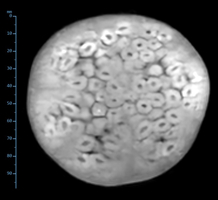
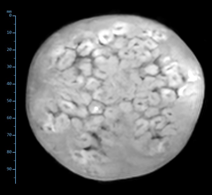
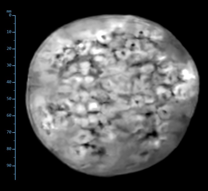
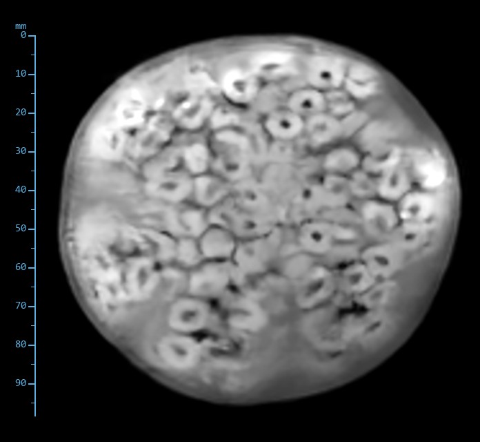
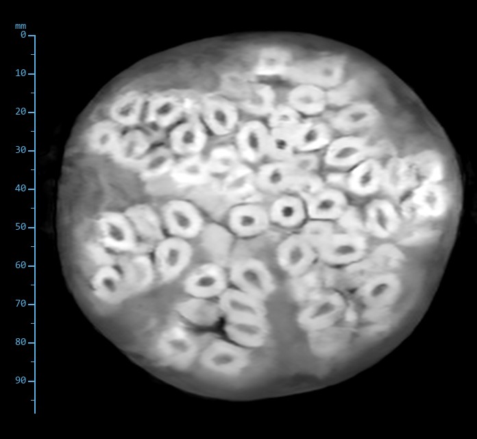
Pomegranate: Water content is not contrast

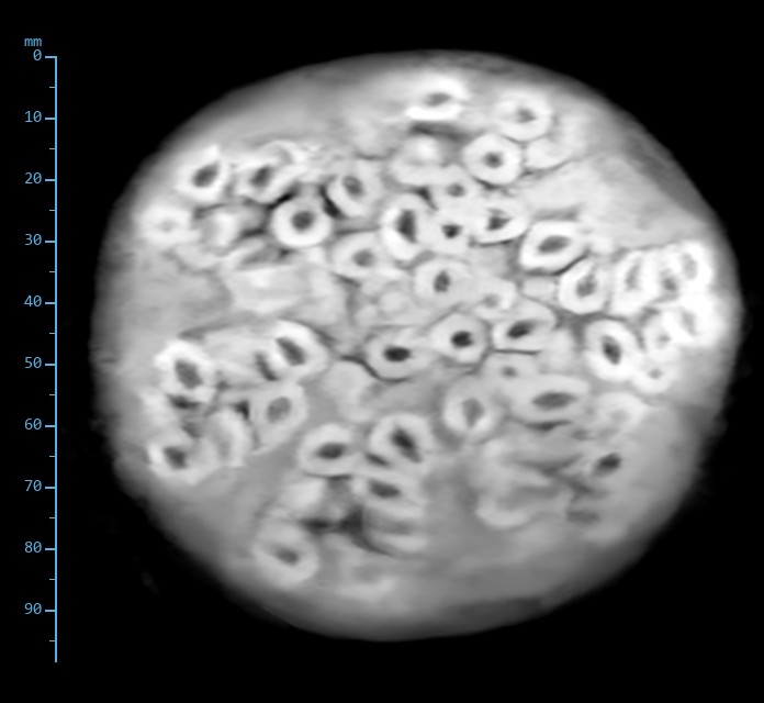
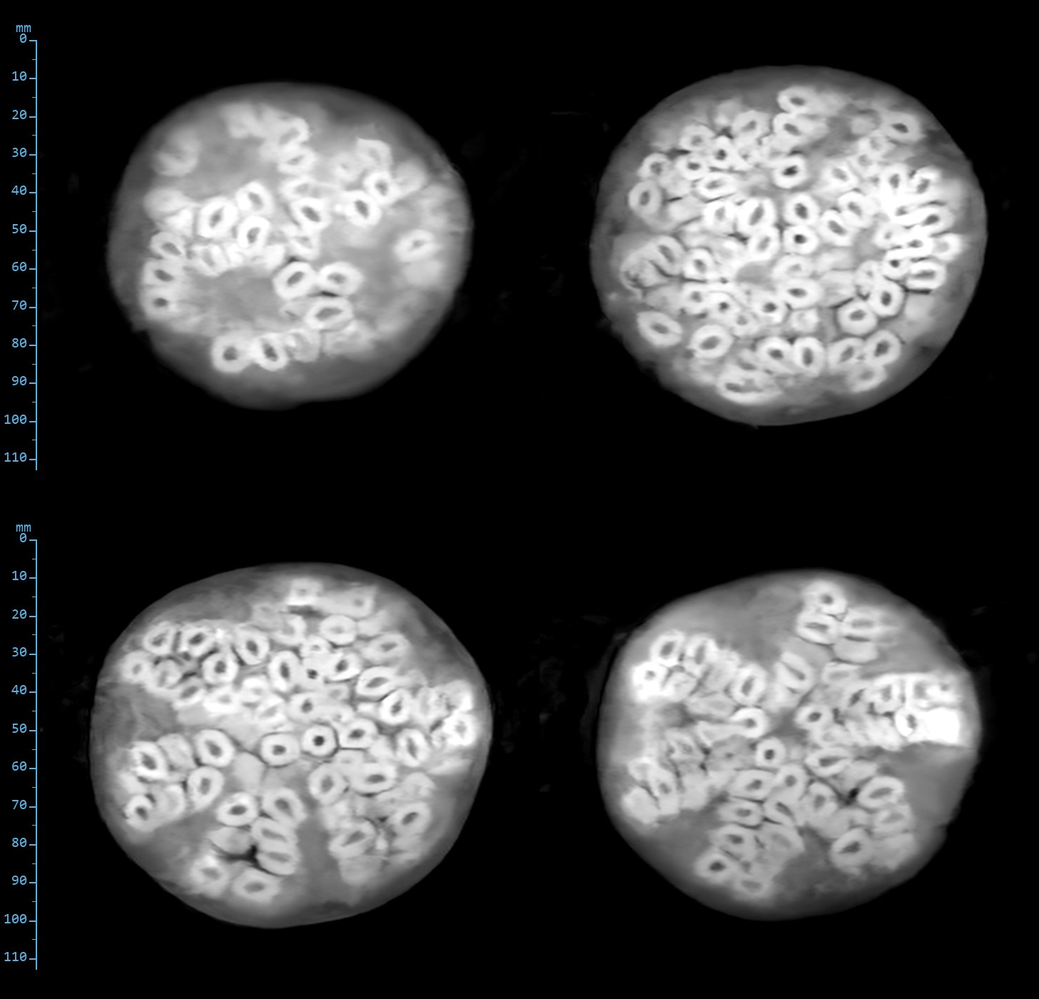
Every part of a pomegranate is wet, so proton density and T1 show almost nothing. On T2 each aril appears as a bright sac of juice around its dark seed, several hundred of them in one slice. Relaxation, not water content, makes the picture.

Slice 10 of 12. Same slice, six sequences.

Proton density spin echo image of the pomegranate
Proton density Spin echoTR 1000 ms · TE 24 ms · 170 × 170, 1.0 × 1.0 × 5 mmNearly flat. The arils and the pith between them have the same proton density.
T1-weighted spin echo image of the pomegranate
T1-weighted Spin echoTR 480 ms · TE 20 ms · 170 × 170, 1.0 × 1.0 × 5 mmStill flat. Short-TR imaging separates nothing here.
T1-weighted gradient echo image of the pomegranate
T1-weighted Gradient echoTR 116 ms · TE 9 ms · 170 × 170, 1.0 × 1.0 × 5 mmFine dark boundaries begin to appear between arils.
T2*-weighted gradient echo image of the pomegranate
T2*-weighted Gradient echoTR 136 ms · TE 9 ms · 170 × 170, 1.0 × 1.0 × 5 mmEach seed is a black dot in its aril; the membranes between clusters show.
T2-weighted fast spin echo image of the pomegranate
T2-weighted Fast spin echoTR 2000 ms · TE 84 ms · 168 × 180, 1.0 × 0.9 × 5 mmJuice bright, seed dark, membranes grey: every aril resolved.
STIR fast spin echo, ti 95 ms image of the pomegranate
STIR Fast spin echo, TI 95 msTR 3160 ms · TE 28 ms · 168 × 180, 1.0 × 0.9 × 5 mmThe same picture with a flatter background. Nothing here contains fat to suppress.
T2-weighted fast spin echo: four slices through the pomegranate
T2-weighted fast spin echo. Slices 6, 8, 10 and 12 of 12. TR 2000 ms, effective TE 84 ms, 168 × 180 acquired, 1.0 × 0.9 × 5 mm.

From home

Duck eggs: Fat, water and cooked protein

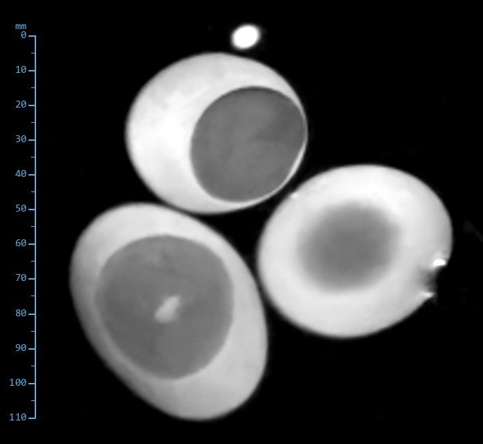
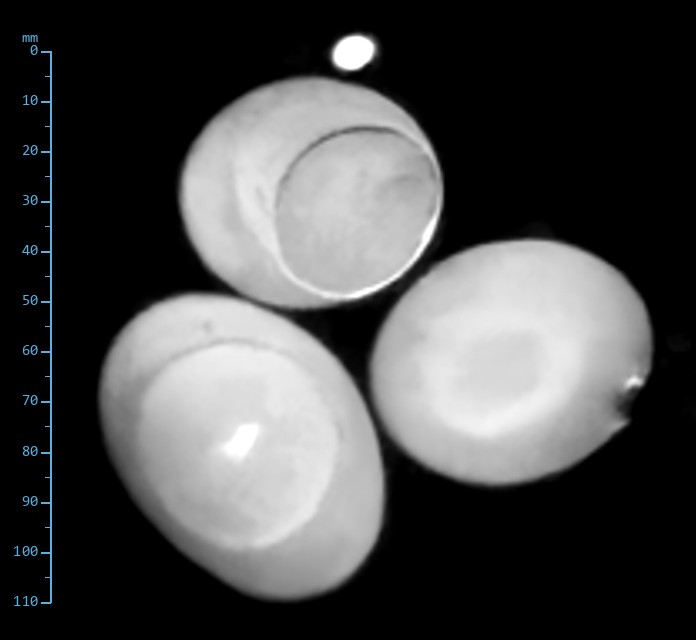
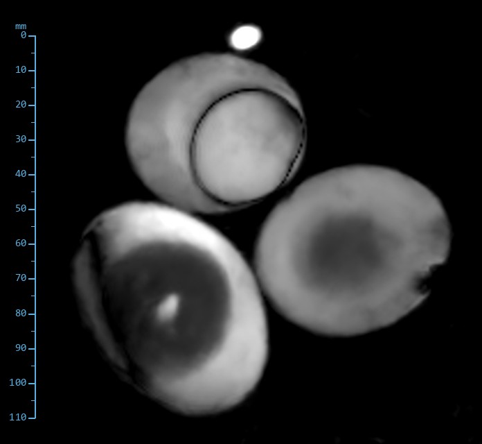
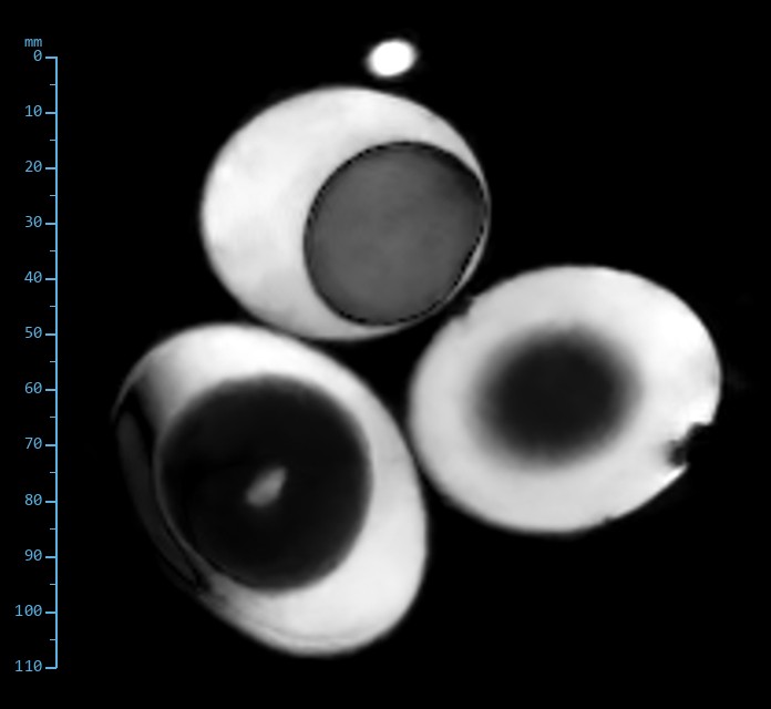
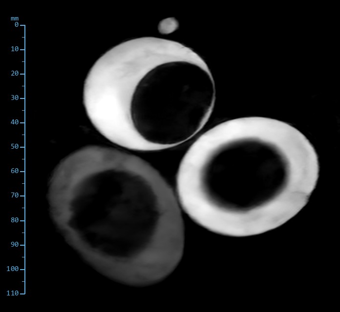
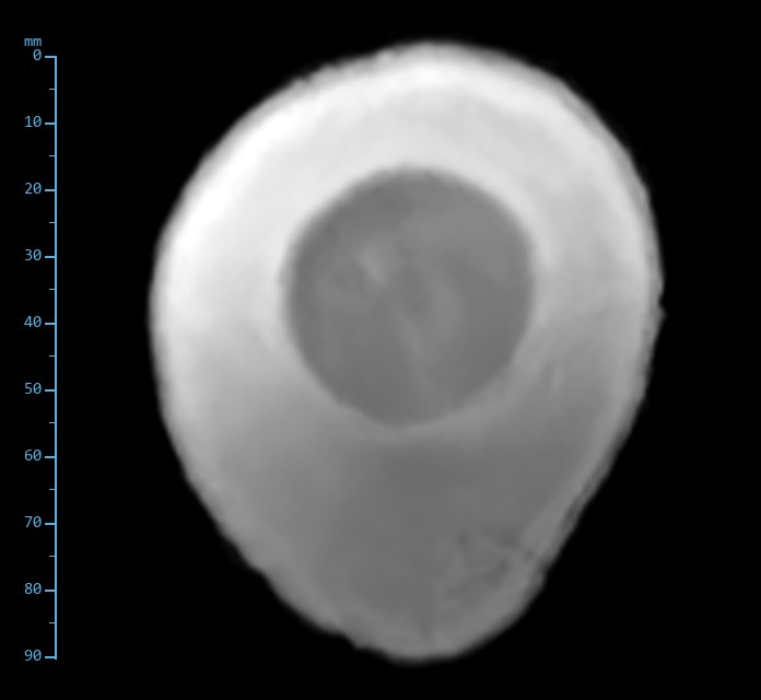
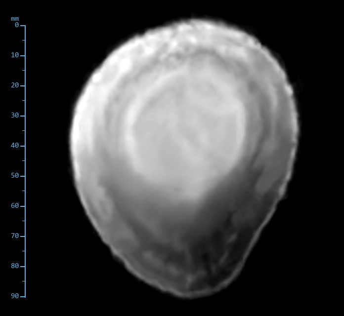
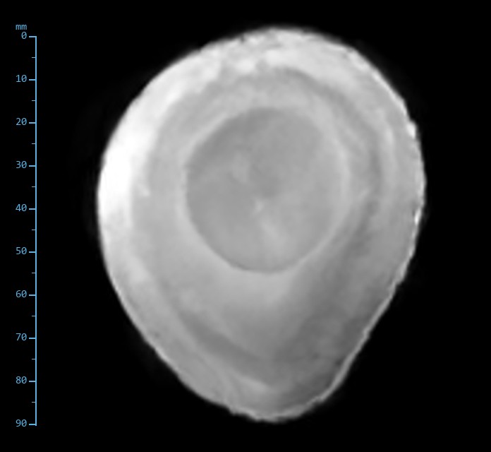
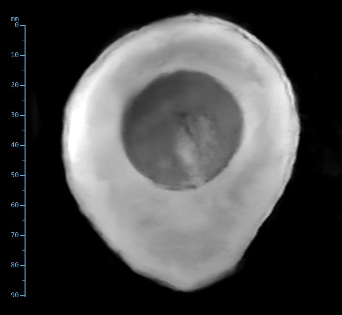
Three duck eggs standing on end: one raw, one hard-boiled, one salt-cured. Two marker tubes identify them. The small tube above the upper-left egg is MCT oil, so that egg is the salted one; the tube to the right of the right-hand egg is water, so that egg is raw; the unmarked egg at lower left is hard-boiled. Albumen is water and protein, yolk is a third lipid, and cooking or curing changes both.

Slice 7 of 12. Same slice, six sequences.

Proton density spin echo image of the duck eggs
Proton density Spin echoTR 1000 ms · TE 24 ms · 170 × 170, 1.0 × 1.0 × 5 mmLiquid whites bright, yolks a shade darker. The salted yolk, firmed by the cure, is a large sharply bounded disc. The cooked white is dim with a brighter rim.
T1-weighted spin echo image of the duck eggs
T1-weighted Spin echoTR 480 ms · TE 20 ms · 170 × 170, 1.0 × 1.0 × 5 mmThe raw yolk brightens against its white, as fat does. The cooked egg is uniformly bright: coagulation shortens T1 as well as T2. The oil marker is bright, the water marker dark.
T1-weighted gradient echo image of the duck eggs
T1-weighted Gradient echoTR 116 ms · TE 9 ms · 170 × 170, 1.0 × 1.0 × 5 mmYolk membranes draw sharp lines. The cooked white is a bright rim around a duller, solid yolk.
T2*-weighted gradient echo image of the duck eggs
T2*-weighted Gradient echoTR 136 ms · TE 9 ms · 170 × 170, 1.0 × 1.0 × 5 mmEvery yolk black. The oil marker stays bright, the cooked white falls away.
T2-weighted fast spin echo image of the duck eggs
T2-weighted Fast spin echoTR 2000 ms · TE 84 ms · 168 × 180, 1.0 × 0.9 × 5 mmRaw and salted whites brightest, cooked white dim, every yolk dark. Protein coagulation shortens T2. The water marker is bright, the oil marker faint.
STIR fast spin echo, ti 95 ms image of the duck eggs
STIR Fast spin echo, TI 95 msTR 3160 ms · TE 28 ms · 168 × 180, 1.0 × 0.9 × 5 mmYolks and the oil marker nulled together at the fat inversion time; the water marker and the liquid whites are the brightest objects in the image.
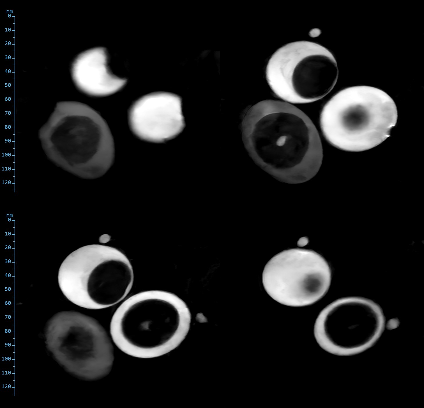
T2-weighted fast spin echo: four slices through the duck eggs
T2-weighted fast spin echo. Four slices through the three eggs. TR 2000 ms, effective TE 84 ms, 168 × 180 acquired, 1.0 × 0.9 × 5 mm.

Produce

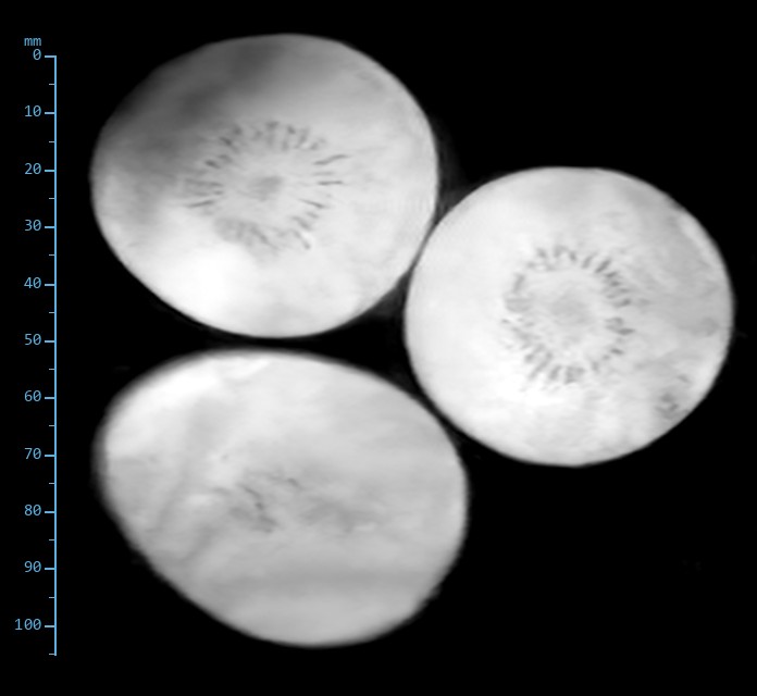
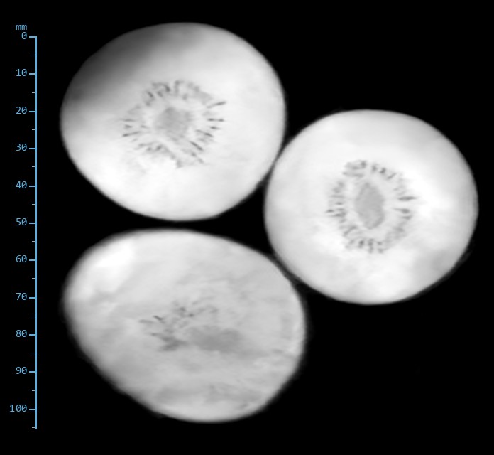
Kiwis: A ring of seeds a millimetre across

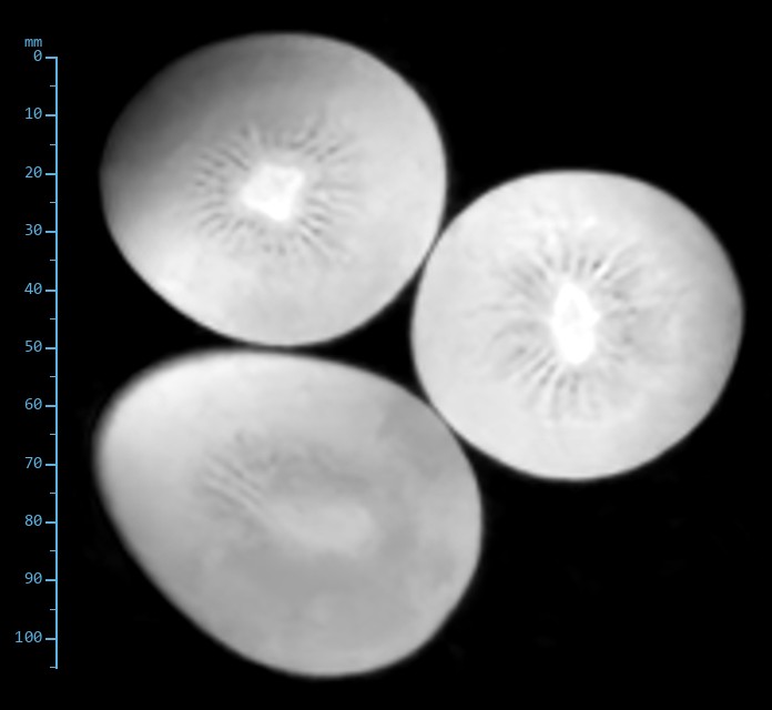
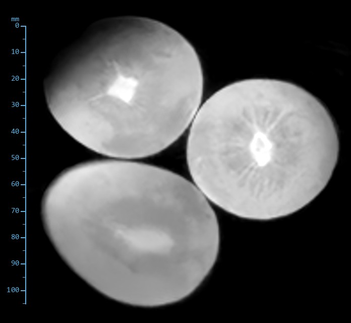
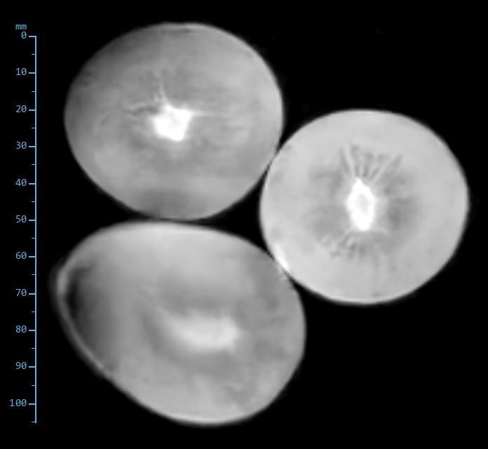
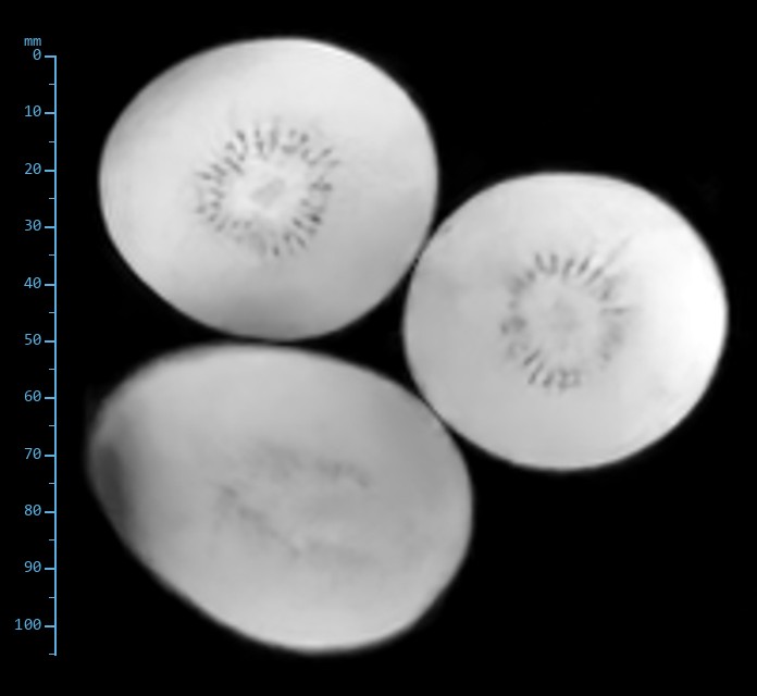
Three kiwis, whole. The flesh is uniform and wet; the interest is the ring of black seeds around the pale core and the fine radial fibres. Where the pomegranate tested contrast, this tests edge resolution on small, dark objects in a bright field.

Slice 9 of 12. Same slice, six sequences.

Proton density spin echo image of the kiwis
Proton density Spin echoTR 1000 ms · TE 24 ms · 170 × 170, 1.0 × 1.0 × 5 mmCore brightest, flesh uniform, the seed ring just visible as a faint dotted circle.
T1-weighted spin echo image of the kiwis
T1-weighted Spin echoTR 480 ms · TE 20 ms · 170 × 170, 1.0 × 1.0 × 5 mmThe core brightens further; seeds are lost in the flesh.
T1-weighted gradient echo image of the kiwis
T1-weighted Gradient echoTR 116 ms · TE 9 ms · 170 × 170, 1.0 × 1.0 × 5 mmRadial fibres from core to skin begin to show.
T2*-weighted gradient echo image of the kiwis
T2*-weighted Gradient echoTR 136 ms · TE 9 ms · 170 × 170, 1.0 × 1.0 × 5 mmEach seed a sharp black dot. Susceptibility makes small dark objects larger and easier to see.
T2-weighted fast spin echo image of the kiwis
T2-weighted Fast spin echoTR 2000 ms · TE 84 ms · 168 × 180, 1.0 × 0.9 × 5 mmSeeds and the radial fibres dark against bright flesh; the core drops out.
STIR fast spin echo, ti 95 ms image of the kiwis
STIR Fast spin echo, TI 95 msTR 3160 ms · TE 28 ms · 168 × 180, 1.0 × 0.9 × 5 mmSame as T2 with the core darker still. Nothing here contains fat.
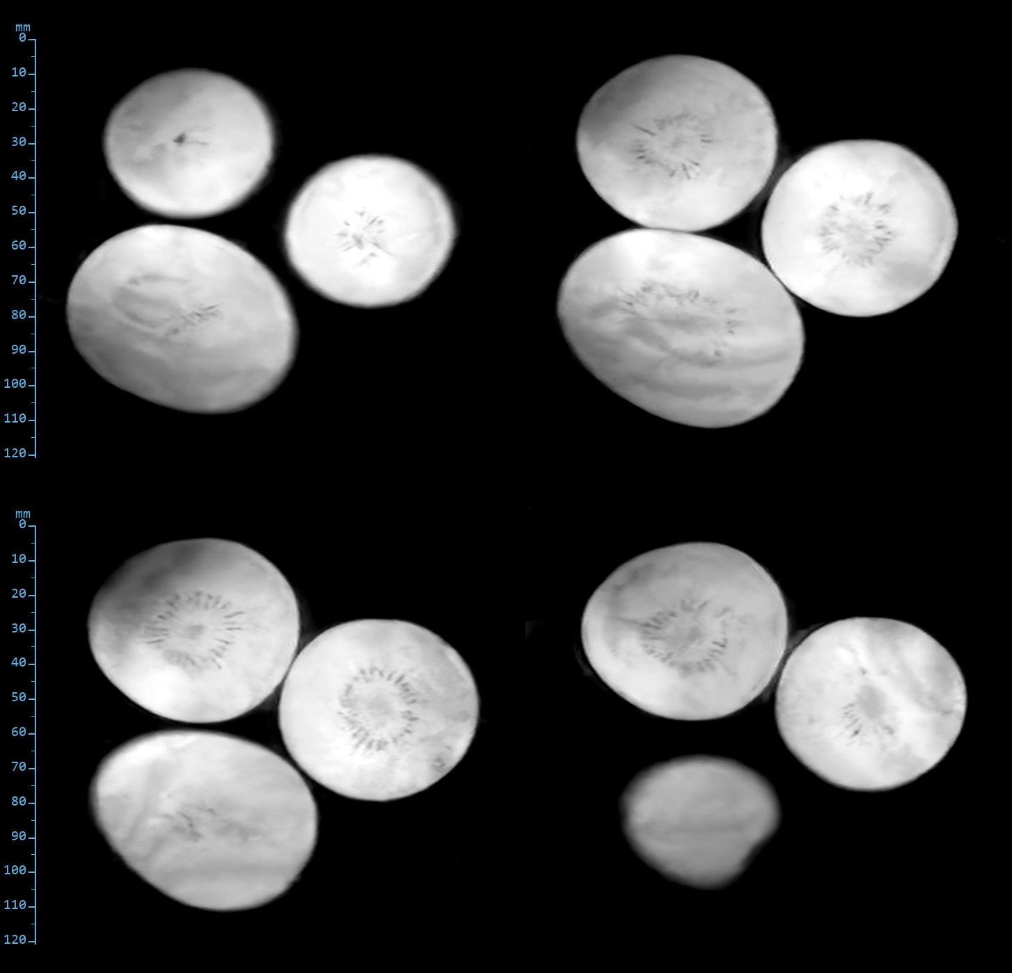
T2-weighted fast spin echo: four slices through the kiwis
T2-weighted fast spin echo. Slices 5, 7, 9 and 11 of 12. TR 2000 ms, effective TE 84 ms, 168 × 180 acquired, 1.0 × 0.9 × 5 mm.

Produce

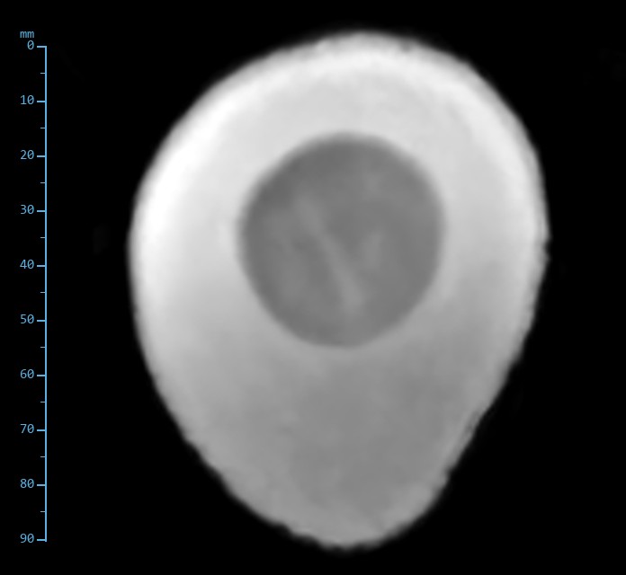
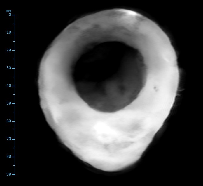
Avocado: A fruit that is partly fat

Avocado flesh is about fifteen percent oil held in a watery pulp, so it sits between the water-only fruit and the yolks. The stone is dry and dense. Ask one question of each image: does the flesh behave like water, like fat, or like a mixture?

Slice 11 of 12. Same slice, six sequences.
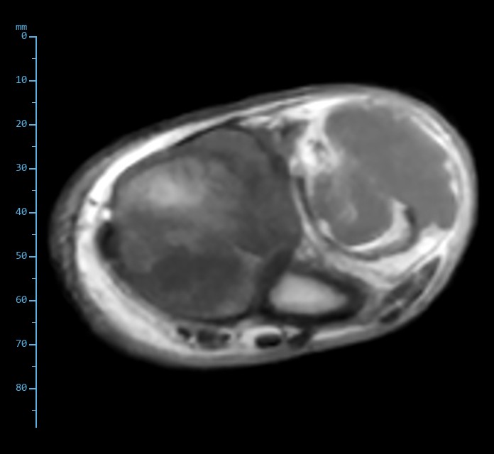
Proton density spin echo image of the avocado
Proton density Spin echoTR 1000 ms · TE 24 ms · 170 × 170, 1.0 × 1.0 × 5 mmFlesh bright and uniform; the stone a slightly darker disc.
T1-weighted spin echo image of the avocado
T1-weighted Spin echoTR 480 ms · TE 20 ms · 170 × 170, 1.0 × 1.0 × 5 mmFlesh brighter than the water-only fruit, as a fat fraction predicts; stone dark.
T1-weighted gradient echo image of the avocado
T1-weighted Gradient echoTR 116 ms · TE 9 ms · 170 × 170, 1.0 × 1.0 × 5 mmFibrous strands through the flesh show as dark lines.
T2*-weighted gradient echo image of the avocado
T2*-weighted Gradient echoTR 136 ms · TE 9 ms · 170 × 170, 1.0 × 1.0 × 5 mmStone dark with a sharp outline, flesh bright.
T2-weighted fast spin echo image of the avocado
T2-weighted Fast spin echoTR 2000 ms · TE 84 ms · 168 × 180, 1.0 × 0.9 × 5 mmFlesh intermediate, stone dark. The oil fraction shortens the T2 of the pulp.
STIR fast spin echo, ti 95 ms image of the avocado
STIR Fast spin echo, TI 95 msTR 3160 ms · TE 28 ms · 168 × 180, 1.0 × 0.9 × 5 mmThe flesh loses signal relative to T2 but does not null: an oil-in-water emulsion is not fat. The stone stays dark.
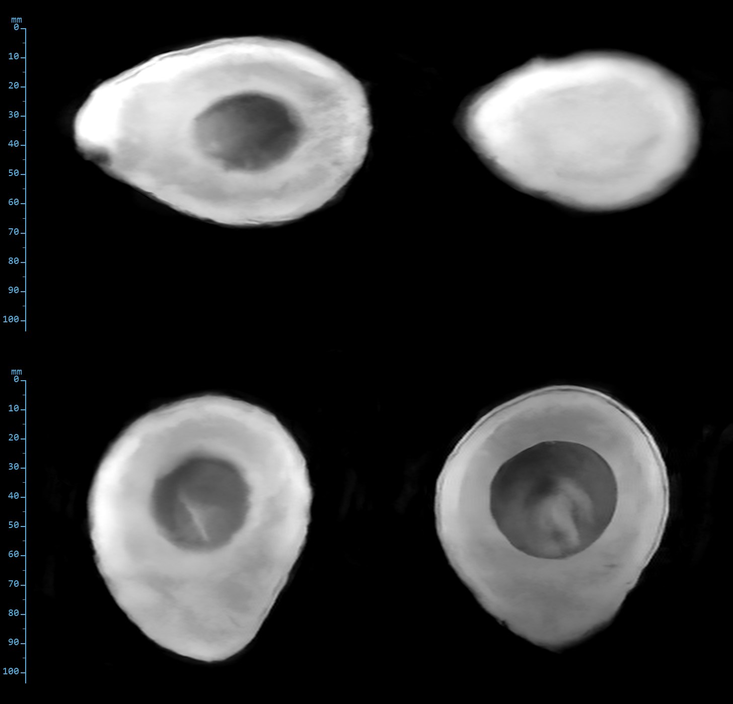
T2-weighted fast spin echo: four slices through the avocado
T2-weighted fast spin echo. Slices 2, 4, 10 and 12 of 12, through the stone twice. TR 2000 ms, effective TE 84 ms, 168 × 180 acquired, 1.0 × 0.9 × 5 mm.

Produce

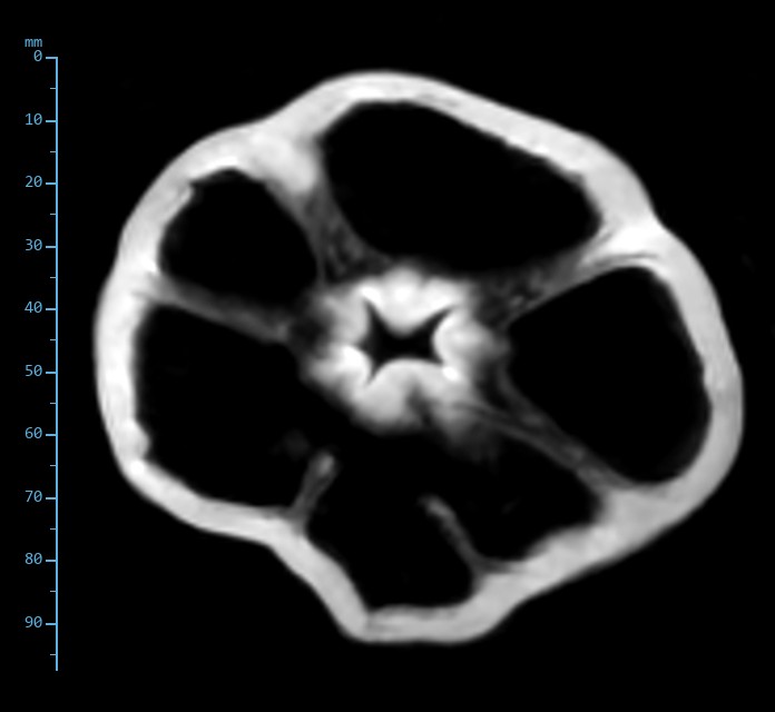
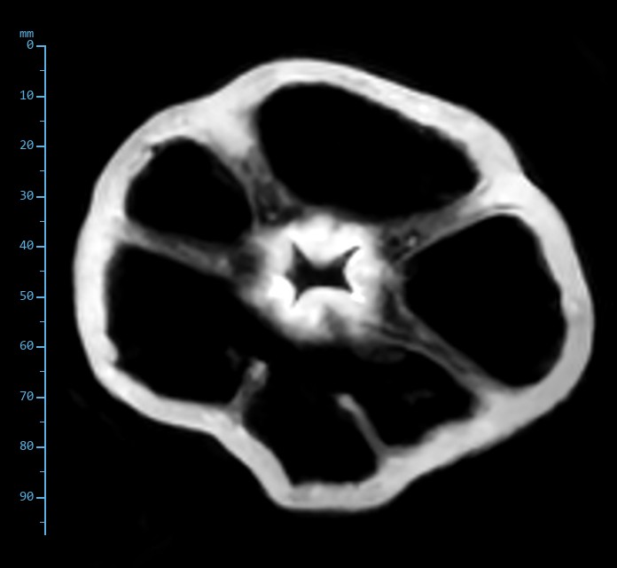
Bell pepper: Mostly air

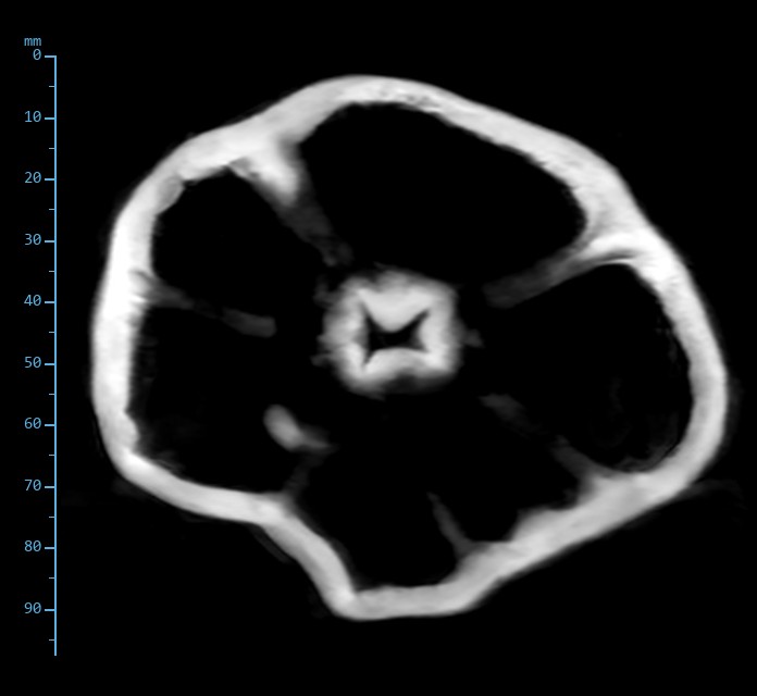
A pepper is a thin wall of wet tissue around air, with a placenta and seeds at the centre. The wall is bright on everything. The test is the air interface: at high field the susceptibility step at air blooms on gradient echo and distorts the wall; at 0.4 T the step is eight times smaller.

Slice 9 of 12. Same slice, six sequences.
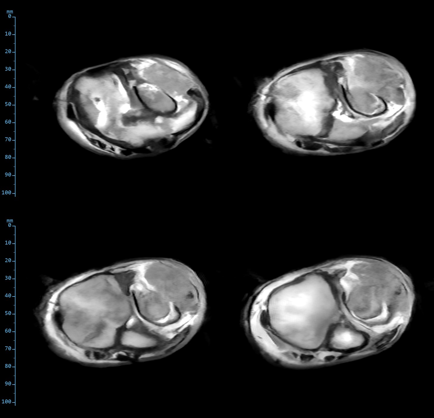
Proton density spin echo image of the bell pepper
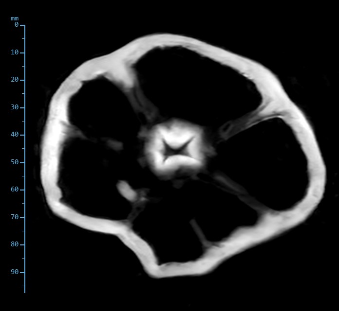
Proton density Spin echoTR 1000 ms · TE 24 ms · 170 × 170, 1.0 × 1.0 × 5 mmWall, septa and placenta bright; the lobes black.
T1-weighted spin echo image of the bell pepper
T1-weighted Spin echoTR 480 ms · TE 20 ms · 170 × 170, 1.0 × 1.0 × 5 mmSame anatomy, placenta brightest.
T1-weighted gradient echo image of the bell pepper
T1-weighted Gradient echoTR 116 ms · TE 9 ms · 170 × 170, 1.0 × 1.0 × 5 mmGradient echo: the wall is as sharp as on spin echo. No blooming at the air interface.
T2*-weighted gradient echo image of the bell pepper
T2*-weighted Gradient echoTR 136 ms · TE 9 ms · 170 × 170, 1.0 × 1.0 × 5 mmStill sharp. The placenta is a bright star at the centre; the air around it draws no halo.
T2-weighted fast spin echo image of the bell pepper
T2-weighted Fast spin echoTR 2000 ms · TE 84 ms · 168 × 180, 1.0 × 0.9 × 5 mmWall and placenta bright. The lobes are air and give no signal at any echo time.
STIR fast spin echo, ti 95 ms image of the bell pepper
STIR Fast spin echo, TI 95 msTR 3160 ms · TE 28 ms · 168 × 180, 1.0 × 0.9 × 5 mmUnchanged. There is no fat in a pepper.
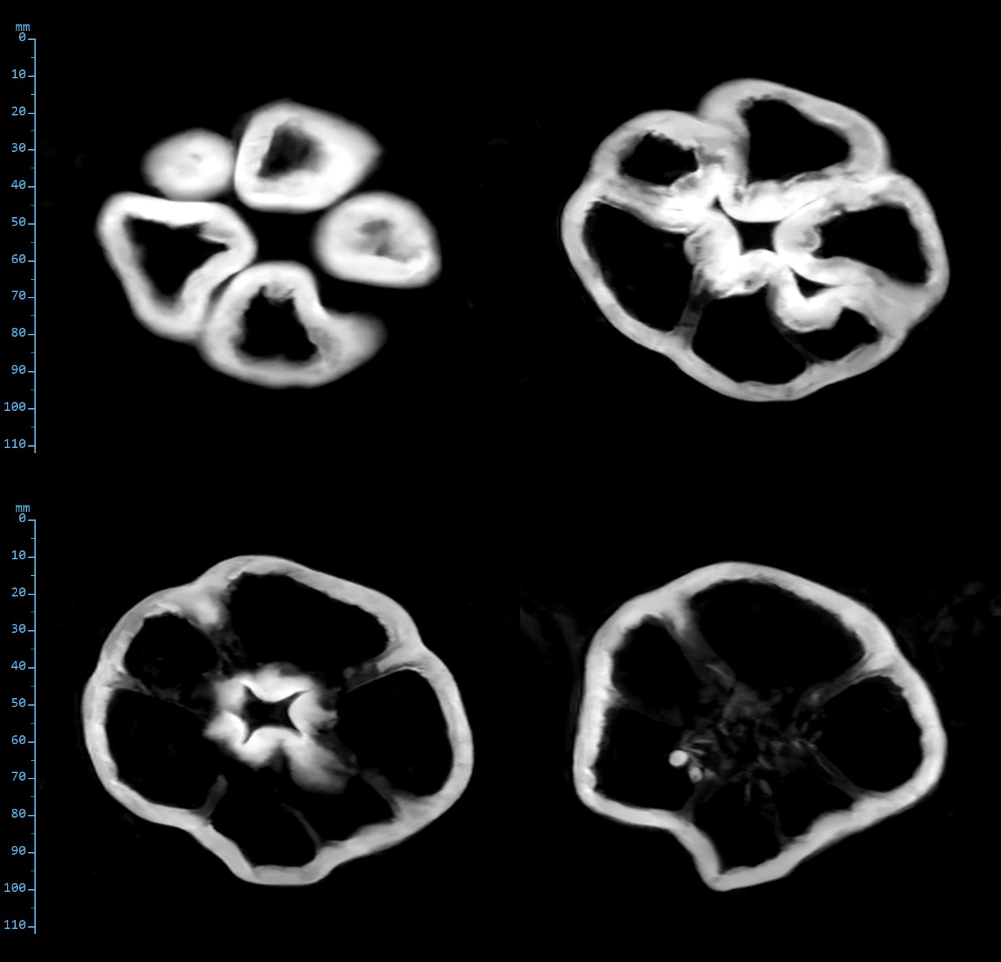
T2-weighted fast spin echo: four slices through the bell pepper
T2-weighted fast spin echo. Slices 4, 6, 8 and 10 of 12, shoulder to base. TR 2000 ms, effective TE 84 ms, 168 × 180 acquired, 1.0 × 0.9 × 5 mm.

Dialling the null

Four inversion times, one slice

The protocol STIR uses TI 95 ms. The same slice of the hock was run again at 70, 120 and 135 ms. Subcutaneous fat is the thing to watch, then the boundaries.
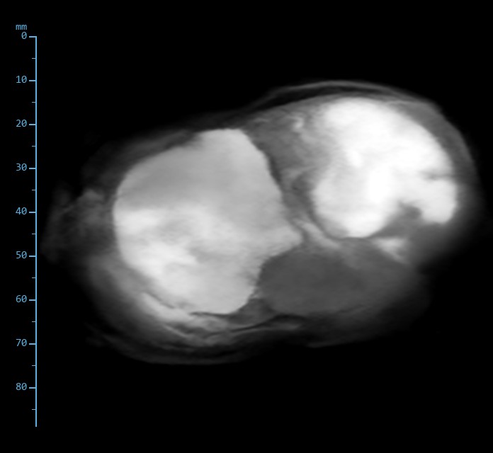
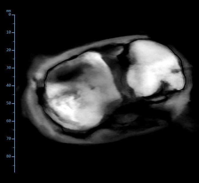
STIR image of the pork hock at inversion time 70 ms
TI 70 msTR 2860 ms · TE 28 ms · 168 × 180, 1.0 × 0.9 × 5 mm · slice 11Too short. Fat has not reached its null; in a magnitude image its inverted signal is still bright and the picture looks like a soft T2.
STIR image of the pork hock at inversion time 95 ms
TI 95 msTR 3160 ms · TE 28 ms · 168 × 180, 1.0 × 0.9 × 5 mm · slice 11The protocol value. Fat at its darkest, no boundary artefact. Water-rich cartilage brightest.
STIR image of the pork hock at inversion time 120 ms
TI 120 msTR 3460 ms · TE 28 ms · 168 × 180, 1.0 × 0.9 × 5 mm · slice 11Just past the null. Fat has crossed zero and now cancels against still-inverted water in partial-volume pixels, drawing a fine black line along every fat boundary.
STIR image of the pork hock at inversion time 135 ms
TI 135 msTR 3640 ms · TE 28 ms · 168 × 180, 1.0 × 0.9 × 5 mm · slice 11Further past. The boundary lines thicken and fat begins to return. This is the India-ink look of an over-long TI.

The fat null for animal fat at 0.4 T sits between 95 and 120 ms, and the right side to err on is short: a slightly short TI costs a little fat suppression, a slightly long one costs edges. The protocol value of 95 ms, chosen on MCT oil in the phantom, holds on the real thing.

What it shows

Three things to take from the shopping

Real tissue behaves

Skin, fat, muscle, tendon, cartilage and cortical bone rank on every sequence exactly as the phantom media predicted they would, and the yolks and avocado flesh behave as the fat fractions they carry.

The null is right

Fat in the hock and lipid in the yolks suppress at the protocol inversion time, with the water marker and the fluid-rich tissues left bright.

Air draws no artefact

A pepper is mostly air. At 0.4 T the air-to-wall interface stays sharp even on gradient echo, where a high-field magnet would bloom. That matters in a hoof.

Reconstructions from the AltraMRI console, 8 and 9 November 2026, windowed per image; standard reconstruction, no further processing. Matrices are as acquired: readout oversampling and navigator echoes are not counted. The grapefruit was re-acquired on 9 November with the fast spin echo readout at the standard 168 samples; the fast spin echo and STIR images of the other objects, from 8 November, were run with 336 readout samples over the same field of view (0.5 × 0.9 mm nominal) and will be re-acquired. The scale on each image is in millimetres, from the acquisition field of view. Objects bought that morning in Littleton, Massachusetts; the eggs from our own ducks. No horse has been imaged on the 0.4T system yet.

Back to the phantom images